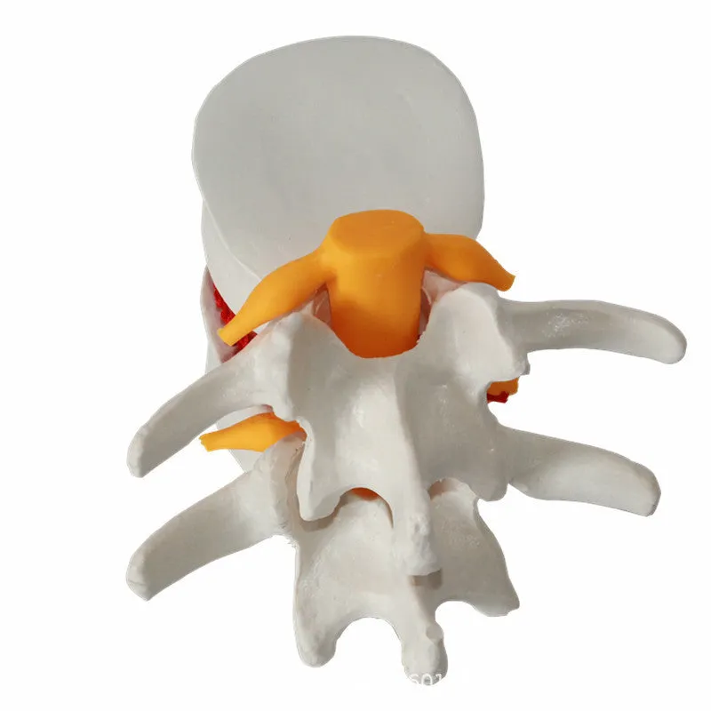
Medical educational demonstration model of human lumbar intervertebral disc herniation, anatomical training aid showing disc pathology, white PVC construction with 11×10×7.6 cm dimensions for teaching spinal conditions in medical schools and clinics.


Medical educational demonstration model of human lumbar intervertebral disc herniation, anatomical training aid showing disc pathology, white PVC construction with 11×10×7.6 cm dimensions for teaching spinal conditions in medical schools and clinics.
Lumbar Intervertebral Disc Herniation Demonstration Model for Medical Education and Clinical Training

This anatomical demonstration model provides healthcare educators, medical students, and clinical professionals with a tangible, three-dimensional representation of lumbar intervertebral disc herniation. Designed specifically for educational purposes, the model illustrates the physical characteristics of this common spinal condition without requiring patient involvement for visual explanation. Made from durable white PVC material and measuring 11×10×7.6 cm, it offers a compact yet detailed reference tool suitable for classroom demonstrations, clinical teaching sessions, or individual study. The model serves as an effective bridge between textbook diagrams and actual clinical observations, helping students and trainees understand the spatial relationships and anatomical changes associated with disc herniation.
Features and Construction
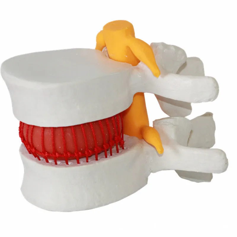
This educational model combines accurate anatomical representation with practical construction for repeated use in training environments. The design focuses on clarity of presentation while maintaining durability suitable for classroom and clinical settings.
Material and Build
The model is constructed from white PVC (polyvinyl chloride), a material chosen for its durability and ability to maintain clear visual presentation through repeated handling. PVC provides sufficient rigidity to maintain the model's anatomical shape while being lightweight enough for easy transportation between teaching locations. The material's surface allows for clear differentiation of anatomical structures relevant to disc herniation pathology, making it suitable for both close-up individual study and group demonstrations where visual clarity from various distances is important.
Size and Practical Fit
With dimensions of 11×10×7.6 cm, the model offers a compact form factor that fits easily on demonstration tables, desks, or examination room surfaces. The size balances sufficient detail for educational purposes with practical portability, allowing educators to transport it between classrooms, lecture halls, or clinical teaching areas. The 0.22 kg weight contributes to this portability while providing enough substance for stable placement during demonstrations. The slightly larger packing dimensions of 11.5×11×8 cm ensure secure transport in its supplied packaging.
Uses and Placement

This demonstration model serves multiple educational functions across different healthcare training environments, from formal medical education to clinical practice settings.
Event or Professional Use
In professional medical education settings, the model functions as a valuable teaching aid during lectures on spinal anatomy and pathology. Medical school instructors can use it to demonstrate the physical characteristics of disc herniation alongside radiographic images or diagnostic findings. Clinical educators in hospitals and rehabilitation centres can employ it when explaining spinal conditions to patients or training junior staff members. The model's clear visual representation helps bridge the gap between theoretical knowledge and clinical understanding, particularly when discussing treatment approaches or surgical interventions for lumbar disc pathology.
Everyday Home Use
For medical students and healthcare trainees engaged in independent study, the model provides a physical reference point for understanding textbook descriptions of disc herniation. Its compact size allows for use in home study environments, library study sessions, or small group review meetings. The durable construction means it can withstand regular handling during revision periods or examination preparation without significant wear. The model's visual nature supports different learning styles, particularly for those who benefit from three-dimensional anatomical understanding when studying complex spinal conditions.
Benefits and Buying Value

This demonstration model offers practical educational advantages through its clear presentation of a specific spinal condition and durable construction suitable for repeated use.
Reuse and Low Maintenance
The PVC construction provides durability that supports multiple teaching sessions and student handling over time, reducing the need for frequent replacement. Unlike perishable anatomical specimens or fragile models, this demonstration tool maintains its educational value through repeated use in various teaching environments. The material requires minimal maintenance beyond basic cleaning when needed, and its resistance to typical classroom wear makes it suitable for institutional purchasing where longevity and cost-effectiveness across multiple academic terms are considerations.
Why Choose This Product
This model addresses the specific educational need for a clear, tangible representation of lumbar intervertebral disc herniation without requiring access to clinical cases or complex imaging equipment. Its focused design on a single pathological condition allows for detailed demonstration that more general spinal models might not provide. The combination of durable construction, practical size, and clear visual presentation makes it suitable for both individual and group learning situations across different educational levels, from undergraduate medical education to postgraduate clinical training programmes.